Build innovative biomarkers

Build innovative biomarkers

Build AI-powered CDx customized to the underlying biology to increase success rate for clinical trials.
Reveal deeper, accurate target insights

Reveal deeper, accurate target insights

Precisely quantify and characterize target expression from IHC images—from sub-cellular distribution to whole slide level analyses and cohort level insights.
Identify high-value drug targets

Identify high-value drug targets

Identify drug targets with precision through surfaceome profiling, tumor-specificity, and membrane specificity analysis.

Streamlined, Scalable
IHC Analysis for Every Stage
of Oncology Development

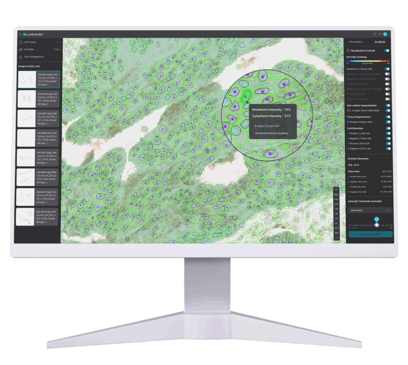
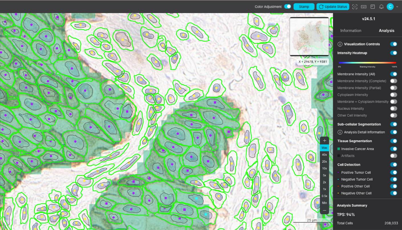
Lunit SCOPE uIHC is an AI-powered digital pathology image analysis software designed to streamline IHC biomarker analysis across various cancer types.

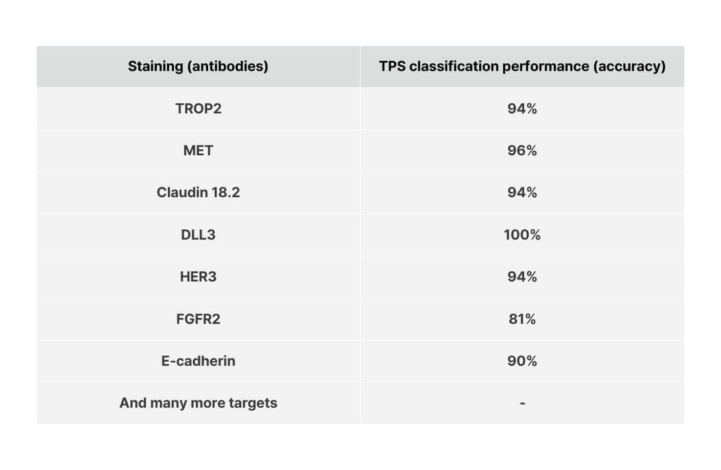
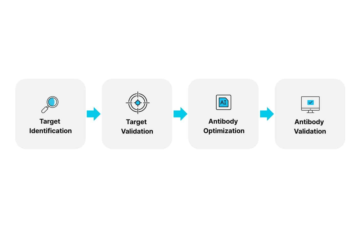
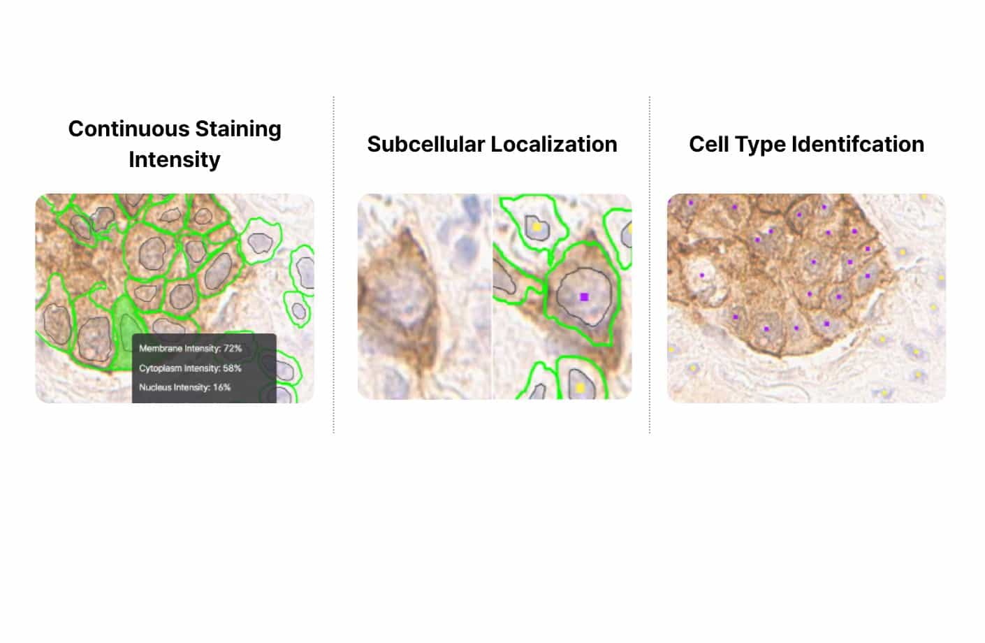
Trained on over 18 IHC stains spanning 20+ primary tumor origins, it supports CDx development for next-gen biomarkers, reveals drug target insights with high membrane and tumor specificity, and drives translational research.

How Lunit SCOPE uIHC Supports Your Team

Table showing IHC biomarker classification performance for antibodies including TROP2, MET, and DLL3, with accuracy rates up to 100%.

Advanced biomarker analysis for translation research

Support comprehensive IHC biomarker analysis—even in early development stages—while capturing a deeper understanding of the target expression on a subcellular, cellular, and whole slide level to inform further advancement of biomarker development.
Digital pathology image showing AI-analyzed tissue with outlined cell membranes and heatmap visualization for next-generation IHC biomarker development.

CDx development for next-gen IHC biomarkers

Use AI-powered IHC analysis—including subcellular localization, continuous scoring, spatial mapping, and ultra-low detection—to develop innovative biomarkers, scoring methods, and cut-offs tailored to your therapy’s mechanism of action.

Target validation and antibody optimization

Identify high tumor and membrane specificity drug targets with scalable IHC analysis. Explore surfaceome profiles, and generate insights that support target selection and therapeutic design.
Workflow diagram showing the AI-driven process from target identification to antibody validation in IHC biomarker research.

Lunit SCOPE uIHC key features

Key features include:
  • Continuous staining intensity quantification (0-100%) for each cell and subcellular components.
  • Subcellular localization of target expression (membrane, cytoplasm, nucleus).
  • Cell type identification (tumor cell, lymphocyte, and other cell).
  • Advanced biomarker analysis integrating quantitative intensity measurement, spatial profiling, and pattern recognition—applied individually or in combination to derive robust insights.
  • Interactive visualizer enabling real-time intensity cut-off assessment.
Microscopic tissue images showing AI quantification of staining intensity, subcellular localization, and cell type identification for IHC analysis.

AI Pathology Software Built to Support Biopharma Innovation

Our digital image analysis software helps biopharma companies scale oncology R&D, advance biomarker strategies, and optimize trials. Lunit is built for end-to-end CDx development, with mature regulatory capabilities, broad scanner and file type compatibility, and scalable deployment across digital pathology platforms and CRO partners.
Breakthrough Insights to Drive Success in Drug Development

Breakthrough Insights to Drive Success in Drug Development

Equip your team with IHC quantification software that accelerates biomarker discovery, enhances CDx strategies, and enables scalable deployment.

Frequently Asked Questions

What differentiates Lunit as a partner in AI-powered image analysis?

Our deep expertise, robust clinical evidence, and partnerships with many CROs and digital pathology platform providers set us apart. We prioritize collaboration and offer versatile solutions that are compatible with a wide range of scanners and IMS.